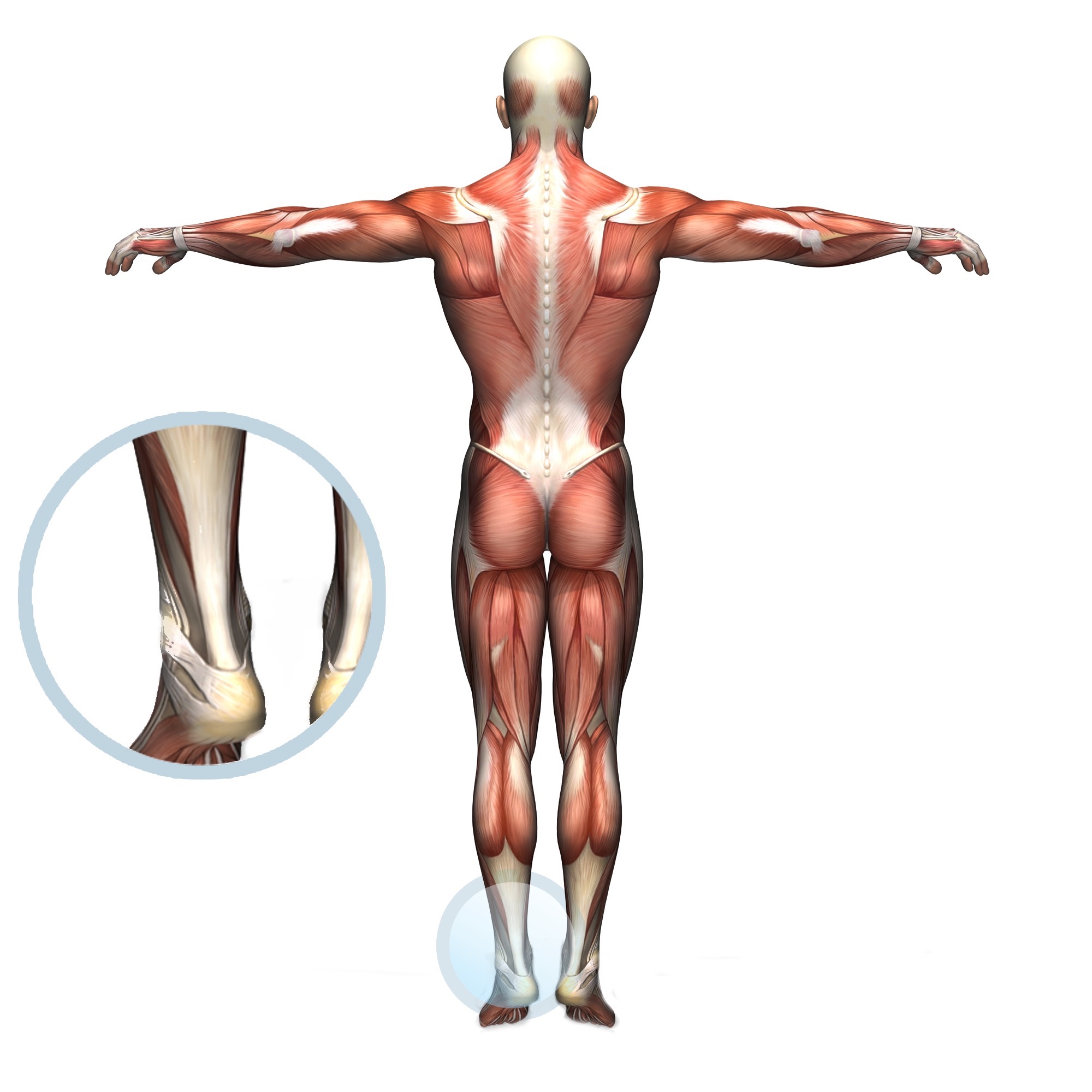
Where is the Achilles tendon?
The location of the Achilles tendon is the bottom part of the calf muscle (technical terms: gastrocnemius, soleus and plantaris muscles), which joins the back of the heel bone (calcaneus).
What is Achilles tendinopathy?
Achilles tendinopathy occurs through irritation of the tendon sheath, or paratendon, which can cause pain and swelling on the back of the heel. This is a common cause in athletes, runners and hikers1,2. The majority of Achilles tendinopathy cases occur in males, typically in 15% of recreational runners, and is the cause of 24% of life-time injuries in athletes. However, a third of chronic Achilles tendinopathy cases are reported in less physically-active people3. In addition, 30% of people who have Achilles tendinopathy undergo surgery3.
How does Achilles tendinopathy happen?
There are two main causes of Achilles tendinopathy: the intrinsic (internal to the body) or the extrinsic (from the environment or outside of the body)3. Sometimes it is a combination of both4.
Intrinsic factors include previous injury to the Achilles (which can lead to degeneration of the tendon), gene variations, male hormonal imbalance3, obesity, diabetes, and age can also lead to degeneration of the tendon if not active3,4. Peri-tendinopathy (which affects the tissues surrounding the Achilles tendon) can lead to scar tissue formation in the tendon4.
Extrinsic factors include weight-bearing on hard, slippery or uneven surfaces, poor training regimes, bad footwear, certain medications (for example, fluoroquinolones, which are medicines that kill bacteria)3,6 or hyperpronation of the ankle (where the heel is rolled inwards and the little toe is raised from the floor)4.
Research has shown that hyperpronation or pronation of the ankle can change the synchrony between the joints of the ankle and foot, and therefore the tendon may be put under intense pressure, causing a ‘wringing effect’. The wringing effect can decrease blood flow to the tendon and the tissues around the tendon (paratendon)3. This can lead to possible degeneration of the tendon in the future3.
Underlying mechanisms and theories of Achilles tendinopathy
An Achilles tendinopathy would change the tendon’s appearance, cell structure, and types and numbers of cells. Research suggests that an Achilles tendinopathy would appear to look more yellow/brown when looking through the microscope, compared to that of a healthy tendon, which would look white4. Collagen in the tendon would be disrupted by increasing weaker collagen being promoted in the area that occurs when an injury is present. There can be a decreased number of cells in some areas of the tendon itself4.
Peri-tendinous tissue (tissues around the tendon) in chronic Achilles tendinopathy has shown two major types of cell: myofibroblasts and fibroblasts. Both of these cells help with repair and re-modelling of the tendon, and the addition of contractile cells4. However, the myofibroblasts lay down lots of collagen in the peri-tendinous tissues, but these cells have been reputable for shrinking peri-tendinous tissue and scar tissue formation4. Alongside the inflammation of the Achilles tendon, ‘neovascularisation’ occurs, which means the formation of new vessels, which has been thought to affect treatment performance5.
Symptoms of Achilles tendinopathy
Common symptoms include:
- pain in the back of the heel
- pain in the calf muscle half way down the leg
- decreased range of motion in the heel
- walking may cause discomfort
- swelling localised to the ankle (redness may be observed in early stages).
Diagnosis of Achilles tendinopathy
Besides being checked by an Osteopath or a Physiotherapist at Perfect Balance Clinic, Achilles tendinopathy can be officially diagnosed by:
- MRI scans
- CT scans5.
- ultrasound, which is becoming a very good diagnosis tool and is meant to be the first method of diagnosis5.
Treatment of Achilles tendinopathy
Methods used by Osteopaths and Physiotherapists at Perfect Balance Clinic consist of:
- soft tissue massage
- articulations of the ankle and foot joints
- myofascial release (relaxing the muscles and increasing circulation) of surrounding tissues
- dry needle/acupuncture
- mobility exercises
- rehabilitation with our specialised trainers and pilates instructors
- specialised exercise prescriptions
- injections of collagenase or saline into the tendon5.
References
- Solomon, L., Warwick, D., Nayagam, S. (2010). Apley’s system of Orthopaedics and Fractures. 9th edition. London: Hodder Arnold, pp.614–615.
- Sarimo, J., Orava, S. (2011). Fascial incision and adhesiolysis combined with radiofrequency microtenotomy in treatment with chronic midportion Achilles tendinopathy. Scandanavian journal of surgery, 100(2), pp.125–129.
- Munteanu, E.S., Barton, J.C. (2011). Lower limb biomechanics during running in individuals with Achilles tendinopathy: a systematic review. BioMed Central, 4(15), pp,1–16.
- Van Sterkenburg, N.M., Van Dijk, N.C. (2011). Mid-portion Achilles tendinopathy: why painful? An evidence-based philosophy. Knee Surgery, Sports Traumatology, Arthroscopy, 19(1), pp.1367–1375.
- Wang, Po-H., Luh, J-J., Chen, W-S., Li, M-L. (2011). In vivo photoacoustic micro-imaging of microvascular changes for Achilles tendon injury on a mouse model. Biomedical Optics Express,2(6), pp.1462–1470.
- Durey, A., Baek, Y.S., Park, J.S., Lee, K., Ryu, J-S. (2010). Levofloxacin-induced Achilles tendinitis in a young adult in the absence of predisposing conditions. Yonsei Medical Journal, 51(3), pp.454–456.